Ultra-Low Dose CT KUB
– by Dr. Jeffrey Yeoh, Radiologist –
Stone Follow-Up CT (Ultra-Low Dose CT KUB)
The Ultra-Low Dose CT KUB examination, Noncontrast CT Abdomen and Pelvis exam, is designed specifically for patients who need follow-up imaging of their known urinary tract calculi. The ultra-low dose CT KUB study is expected to be most often requested by urologists, but any physician may also request this service.
Imaging of Urinary Tract Calculi
CT is the “gold standard” in imaging urinary tract calculi. Urinary tract calculi are usually calcified, and therefore best imaged with CT. For calculi over 3 mm CT has reported sensitivity and specificity of nearly 100%. Traditional dose CT can reliably detect urinary tract calculi as small as 1 mm, and can locate them anywhere within the urinary tract, from kidney, ureter, bladder, and even urethra. Abdominal X-ray (KUB) has reported sensitivity of only 70% and is also limited by lack of specificity, since calcified phleboliths and other calcifications often cannot be differentiated from urinary tract calculi. Ultrasound can detect urinary tract calculi, when larger, and in areas well visualized by ultrasound, like the kidney and bladder, but the ureters are usually obscured by bowel gas. Therefore ureteral calculi are more often not seen, even when present.. Ultrasound may detect hydronephrosis and/or hydroureter, which might suggest an unseen obstructing ureteral calculus.
Urinary tract calculi often afflict younger patients, who will likely need many more years of imaging follow up. Frequent imaging of urinary tract calculi is often needed because of the rapid changes in the course of the disease, as the stones may quickly form/dissolve, migrate down or upstream, or possibly obstruct urine flow. Therefore, since this type of imaging follow-up may be needed frequently and in close succession in the same patient, the Ultra-Low Dose CT KUB provides the lowest possible radiation dose that would still allow adequate evaluation of the size, number, and position of the known urinary tract calcified calculi.
Radiation Dose
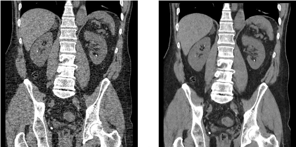
Traditional Noncontrast CT Abdomen and Pelvis protocols effective radiation dose is about 15 mSv. In the interest of limiting radiation to our patients, Hawaii Diagnostic Radiology Services, HDRS, limits the dose to about 5 to 10 mSv. Our brand new GE dual energy CT and VEO supercomputer, one of three in the nation, at our St. Francis Liliha location,utilizes cutting edge technology to improve image quality at ultra-low doses. At this location, we can perform follow up CT KUB exams at about 1-2 mSv. (3-5 mSv in larger patients.)

Left – Low dose image without VEO processing, Right – Low dose image utilizing VEO processing. (Click to enlarge)
Limitations
There is a substantial reduction in the signal to noise of the images generated,) as the CT radiation dose decreases. The detail in the CT images decreases at these ultra-low doses, and therefore the ability to detect, characterize, and evaluate abnormalities other than calcified urinary tract calculi is limited. Consequently, we require a prior “normal dose” CT Abdomen and Pelvis (CT KUB or CT IVP also acceptable) within the prior 6 months before we do this tailored ultra-low dose exam.
The imaging of tiny calcified urinary tract calculi may also be compromised by the lowered radiation dose. The lower the radiation dose, the larger the minimum size calculus detectable. Patient size and body habitus will also determine the minimum dose needed to image adequately. Larger patients require higher radiation techniques to penetrate and visualize their internal anatomy. Initial studies on ultra-low dose CT have shown difficulties in achieving diagnostic exams with ultra-low x-ray dose in patients with body mass index over 30. (BMI = weight (kg) / height (m) squared)
Requirement to Schedule Ultra-Low Dose CT KUB:
Prior (less than 6 months) CT Abdomen/Pelvis. If the prior study was not done at Hawaii Diagnostic Radiology Services, please provide CD of this prior outside CT Abd/Pelvis.
Stone Follow-Up CT Report Disclaimer:
The ultra-low dose CT KUB protocol is designed specifically for calcified urinary tract stone follow-up with the lowest diagnostic radiation dose possible. This exam has limited ability to detect and characterize other potentially significant abnormalities, including but not limited to solid visceral mass lesions. Also, because of the ultra-low radiation dose smaller urinary tract calculi, less than 3 mm in size, may not be seen reliably. Urinary tract stone size may also be over or underestimated by + or – 20% when compared to normal dose techniques.
References:
